
BrainScan
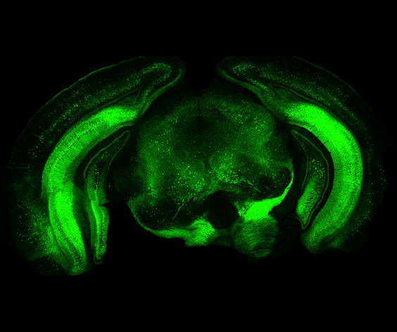
Simplify your neuroscience workflow. BrainScan rapidly scans brain slices to produce high-resolution digital panoramic images, and automatically performs brain region segmentation, atlas registration, and cell counting for quantitative analysis — full automation from imaging to data processing.
Request a Demo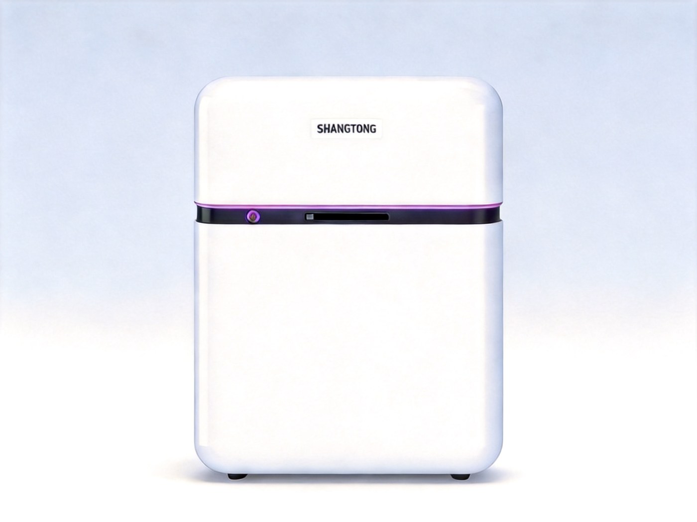
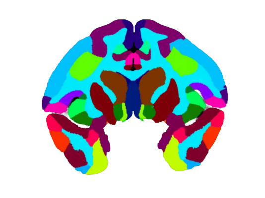
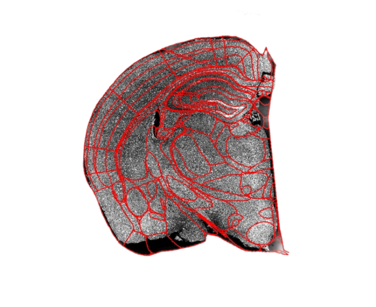
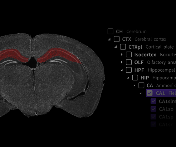
One-Click Atlas Registration
Automatically identifies brain slices and matches them with the Allen Brain Atlas. Supports rat, mouse, macaque and ferret brain atlases.
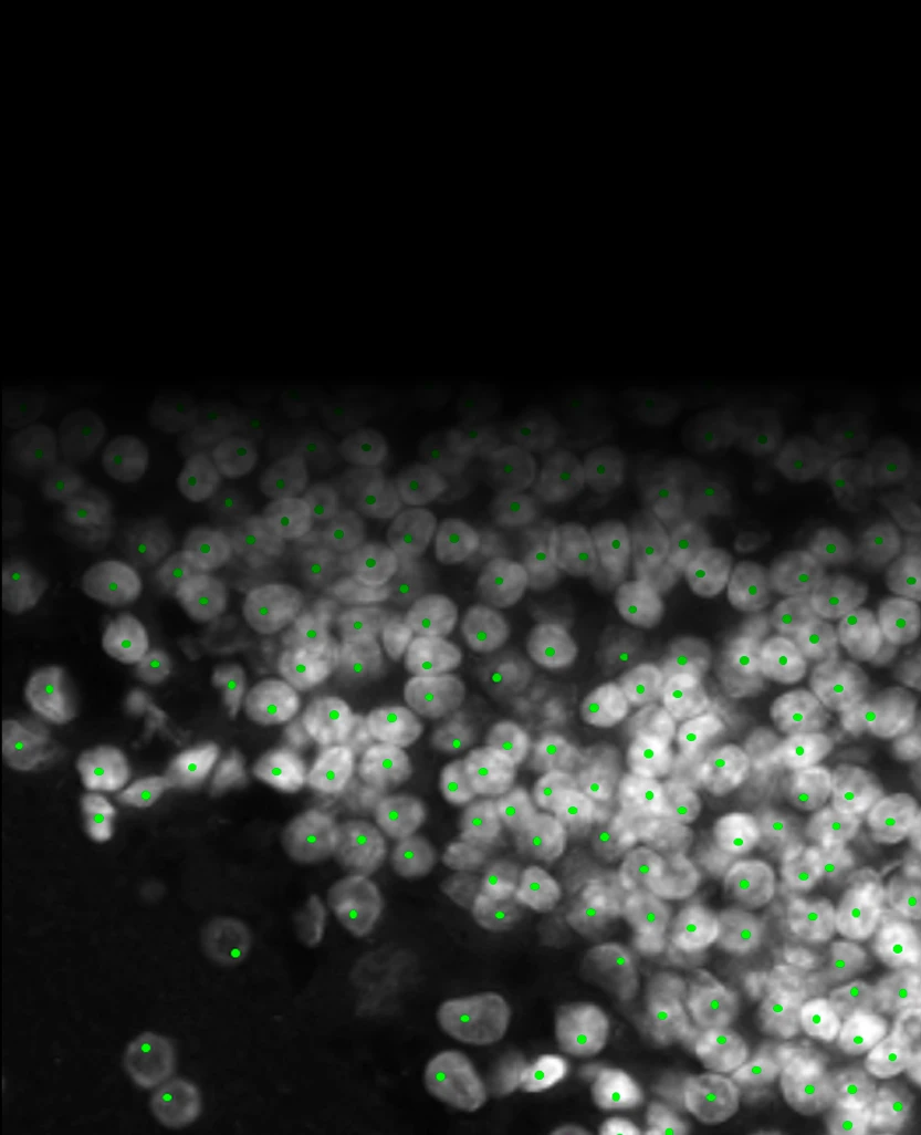
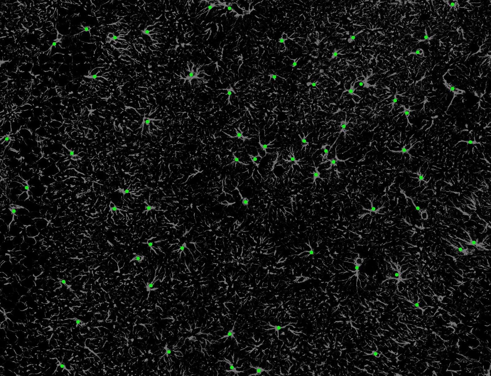
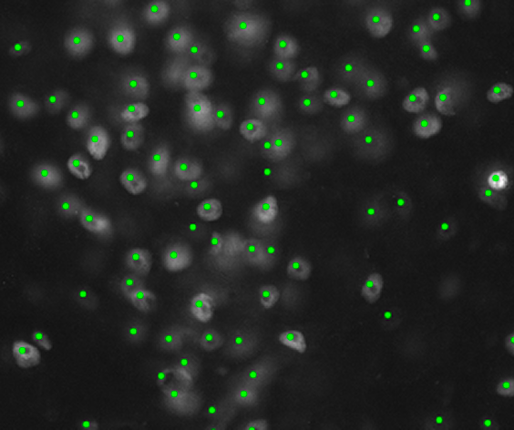
Cell Recognition & Counting
Automatic quantitative analysis that reduces manual intervention, ensuring consistent and accurate experimental results.
Intelligent Scan Path
Automatically identifies ROIs and plans the scanning path — a single brain slice is imaged in under 1 minute (10X, DAPI & GFP, ~40 s).
High-Precision Auto-Focus
Precise focusing on thick brain slice surfaces to quickly capture clear images, keeping all channels on the same focal plane.
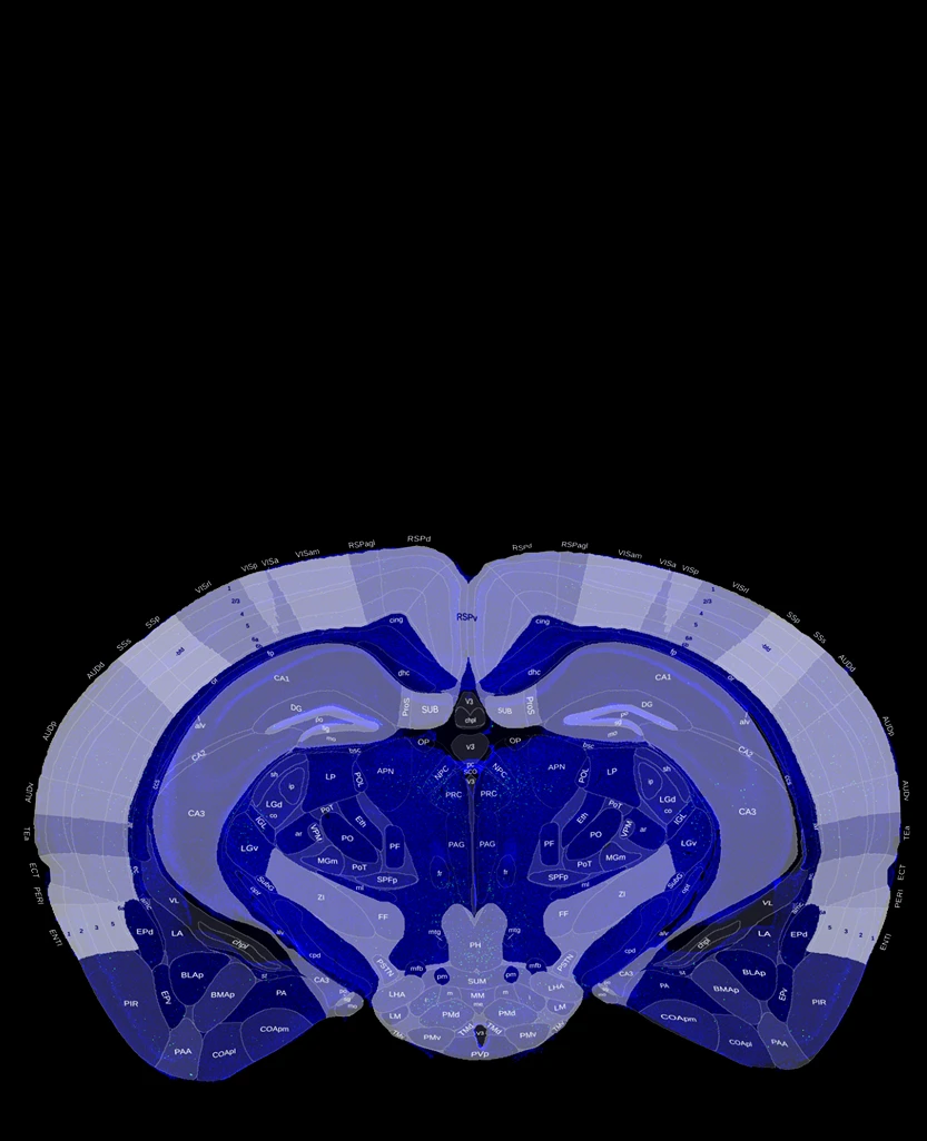
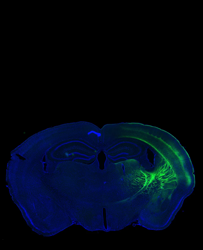
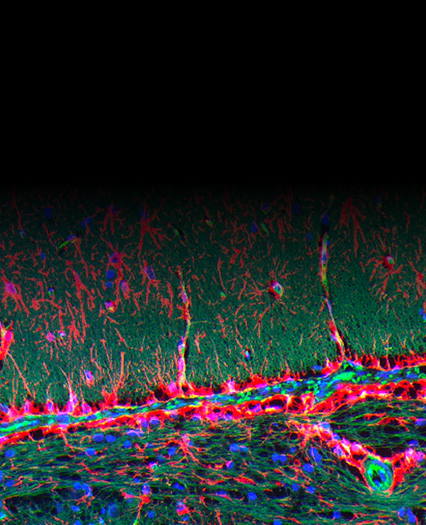
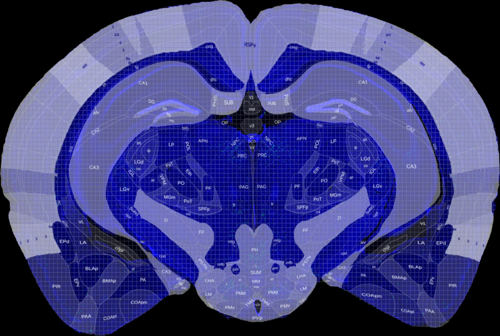
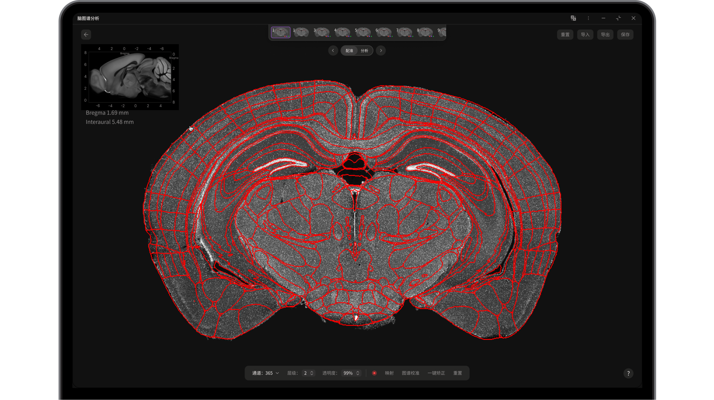
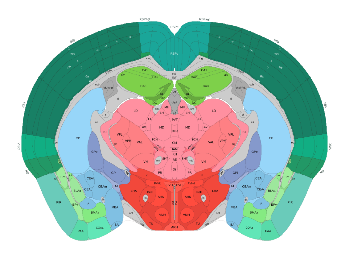
Standard atlas comparison for efficient brain exploration. Register slices automatically and skip the tedious manual work.
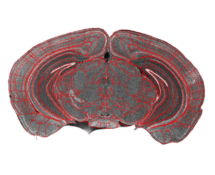
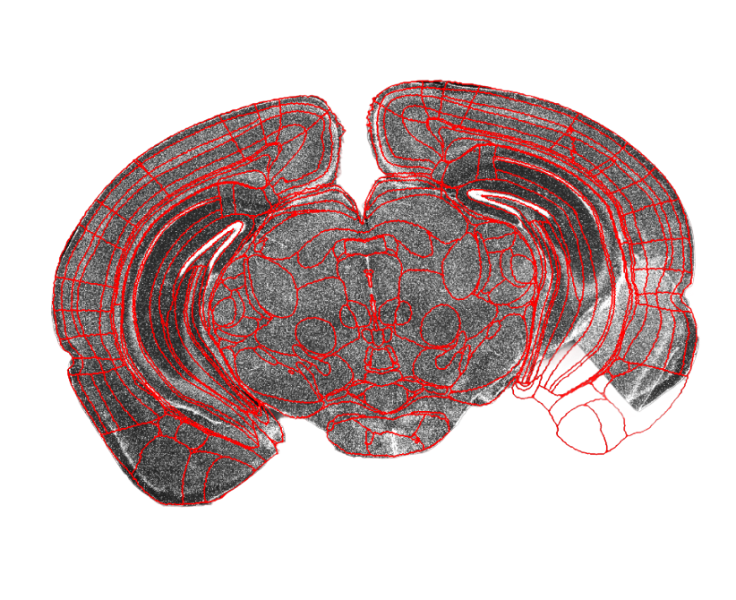
Comprehensive Registration
Handles tilted slices, incomplete slices and half-brain slices with robust algorithms.
- Tilted brain slice registration
- Incomplete brain slice registration
- Half-brain registration
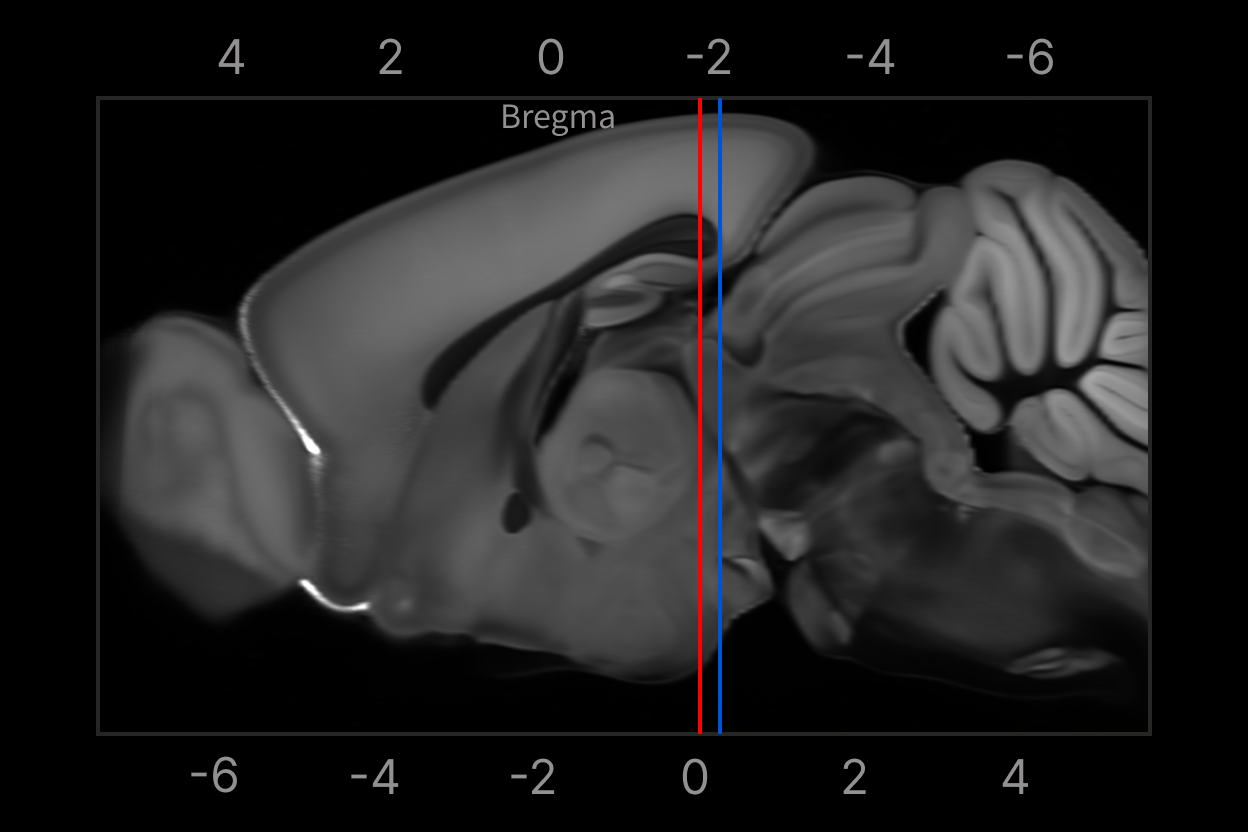
Precise Manual Positioning
Side-by-side standard 2D atlas comparison and Bregma positioning for accurate localization.
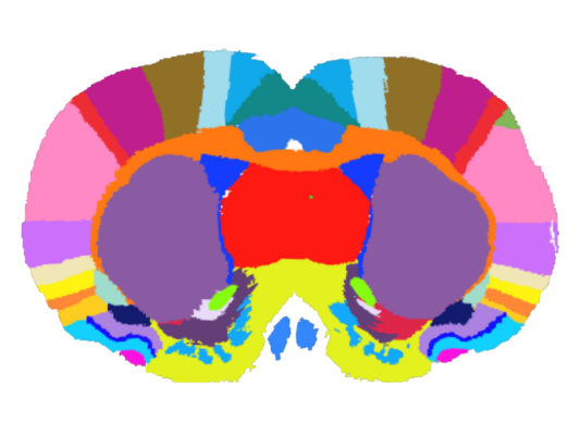
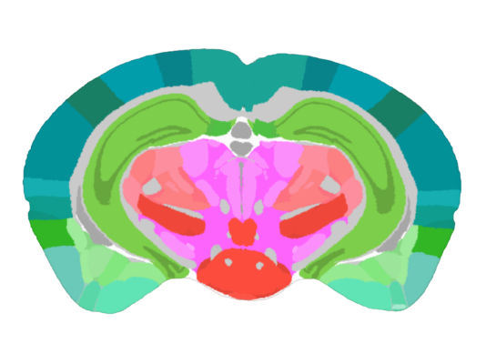

Automatically recognize and quantify signals such as DAPI, c-Fos, neurons and glial cells, with statistics on positive-signal count, area and fluorescence intensity.
Custom Statistical Regions
Supports whole-brain, left/right brain and custom brain region statistics to meet diverse counting needs.
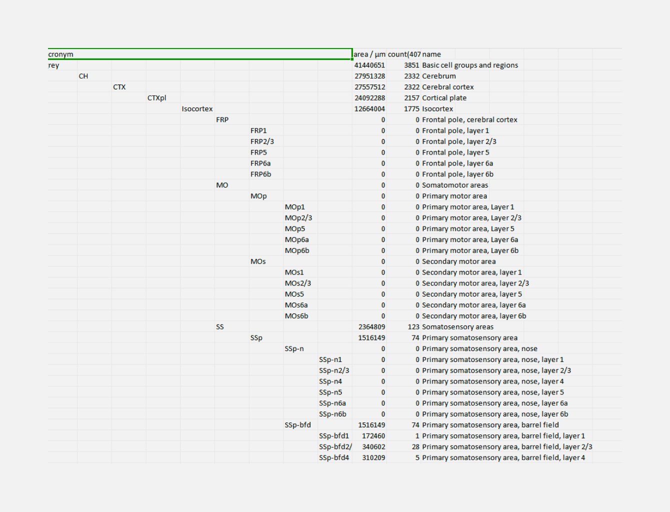
Graded Counting Results
Counting results are presented in a graded Excel layout with precise correlation between parent and sub brain regions.
One Image Per Minute
Automatically recognizes ROIs and plans the scan path — a single slice in under 1 minute.
99% Uniformity
Uniform illumination with global fluorescence background correction achieves up to 99% uniformity.
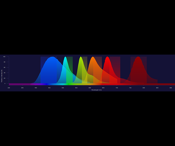
No Crosstalk
Universal fluorescence channel configurations with highly customizable narrow-band filters.
Low Phototoxicity
Strobe mode delivers millisecond high power only during exposure, reducing phototoxicity to samples.
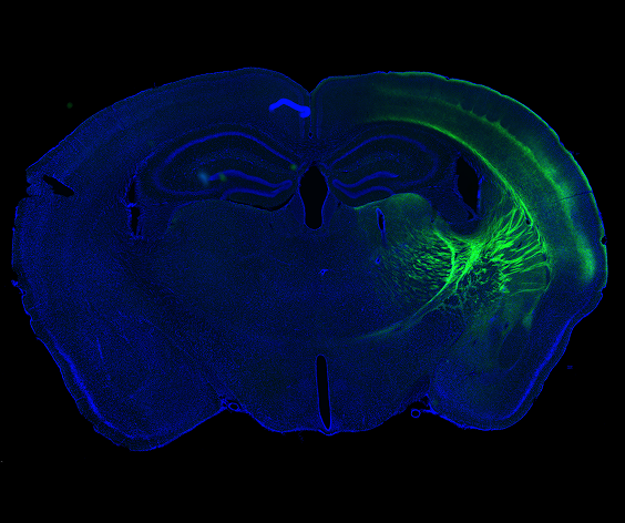
Side LED Illumination
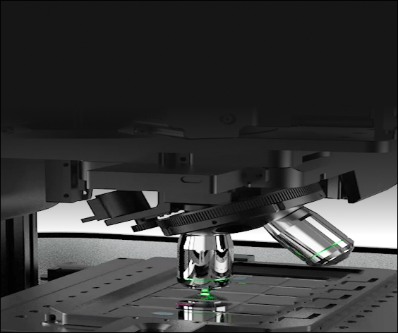
Side LED diffuse illumination delivers high-contrast samples without pre-marking the slide.
Progress Indicator
A ring light on the unit displays scanning status and progress via color.
Parameter Templates
Scan parameters, contrast, analysis regions and export size can be saved as reusable templates.
Batch Export
Export single-channel pseudo-color/gray images or multi-channel overlays in batches.
Bring BrainScan into Your Laboratory
Put brain slice scanning, atlas registration and quantitative analysis into a more efficient automated workflow.
Request a DemoBrainScan Brain Slice Analyzer: High-Resolution Scanning and Automatic Quantitative Analysis
BrainScan is an automated brain slice scanner designed to simplify the neuroscience workflow. It rapidly captures high-resolution digital panoramic images of brain slices and combines atlas registration, brain region segmentation and cell counting in one system, delivering full automation from imaging to data processing. Whether you run a neurobiology laboratory, a pathology facility or a clinical research unit, BrainScan turns tedious manual analysis into reproducible, quantitative results.
Key Features
- Fast whole-slice imaging – acquires complete brain sections as high-resolution digital panoramas in minutes.
- Automatic atlas registration – aligns each slice to a standard brain atlas for consistent region-of-interest mapping.
- Brain region segmentation – automatically delineates anatomical regions for targeted analysis.
- Quantitative cell counting – detects and counts cells with reproducible, unbiased results.
- Low phototoxicity strobe illumination – millisecond high-power exposure minimizes damage to delicate samples.
- Reusable parameter templates – save scan, contrast and analysis settings for standardized workflows.
Technical Specifications
| Imaging Mode | High-resolution widefield scanning, multi-channel fluorescence |
|---|---|
| Scanning Speed | Full brain slice to digital panoramic image in minutes |
| Analysis | Atlas registration, brain region segmentation, automatic cell counting |
| Illumination | Uniform LED with strobe low-phototoxicity mode |
| Export | Single-channel pseudo-color/gray or multi-channel overlays, batch export |
| Usability | Side LED illumination, ring-light progress indicator, parameter templates |
Applications
BrainScan supports a wide range of neuroscience and pathology workflows, including brain slice scanning for cell counting, atlas-based region analysis, neurodegenerative disease research, behavioral neuroscience tissue analysis, and digital pathology in clinical laboratories.
Frequently Asked Questions
What is a brain slice analyzer?
A brain slice analyzer is an automated instrument that scans brain tissue sections into high-resolution digital images and performs quantitative analysis such as atlas registration and cell counting, removing the manual, time-consuming steps of traditional microscopy.
How does BrainScan perform atlas registration?
BrainScan automatically aligns each scanned brain slice to a standard reference atlas, so anatomical regions are consistently identified and compared across samples and experiments.
Can BrainScan count cells automatically?
Yes. BrainScan detects and counts cells automatically with reproducible, unbiased results, supporting both whole-slice and region-specific quantitative analysis.
Which laboratories can use BrainScan?
BrainScan is well suited for neuroscience research laboratories, pathology facilities and clinical research units that need fast, consistent and quantitative brain tissue analysis.
Talk to Our Team About the BrainScan
Tell us about your application — we will confirm specifications, pricing and delivery timeline for your laboratory.
Request a Quote