
LumiFluor AVIS
In Vivo Animal Imaging System
High-sensitivity bioluminescence, fluorescence and chemiluminescence imaging for living small animals — powered by a deep-cooled camera, ring LED excitation and an intelligent gas anesthesia system.
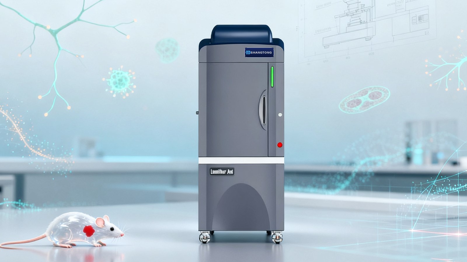
Key Features
Deep-Cooled Imaging
Research-grade camera cooled to -70°C, 7.5 MP output, ≥80% quantum efficiency at 600 nm, and a fast F0.8 lens to capture the faintest in vivo signals.
Multi-Mode Detection
Chemiluminescence, in vivo luminescence and fluorescence — supporting Western/Southern/Northern/Dot blots, luciferase reporters and fluorescent probes.
Precision Optics
Top-mounted ring LED excitation with 22 positions, 18-position emission filter wheel with 8 filters, fully automatic software-controlled switching.
Intelligent Software
Auto-recognition, focus preview, multi-image comparison, pseudo-colour, B-STEP & binning, 3D peak display, ROI selection and auto-exposure.
Core Specifications
| Camera | Deep-cooled research-grade camera |
|---|---|
| Lens | Minimum F value 0.80 |
| Cooling Temperature | -70 °C |
| Effective Pixels | 7.5 million |
| Quantum Efficiency | ≥80% @ 600 nm |
| Pixel Depth | 16 bit |
| Imaging Area | 20 cm × 20 cm |
| Mouse Capacity | 5 mice |
| Temperature-controlled Stage | 20 °C – 40 °C |
| Excitation Source | 22 positions, 22 LED sources included |
| Filter Wheel | 18 positions, 8 emission filters included |
| Dark Box | Fully sealed, side-opening, dual magnetic design |
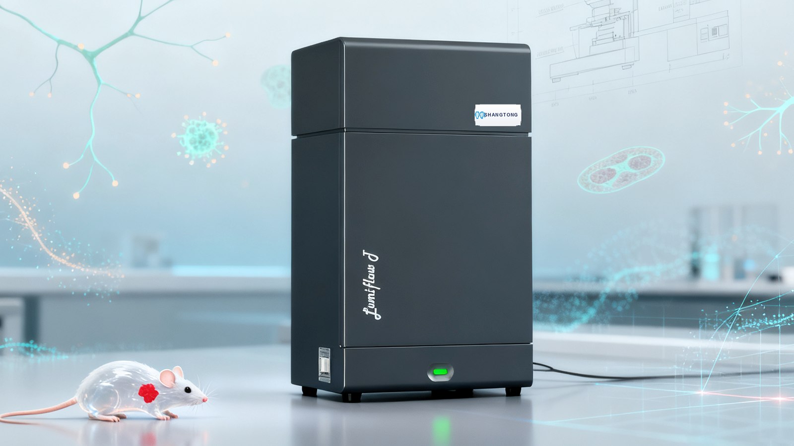
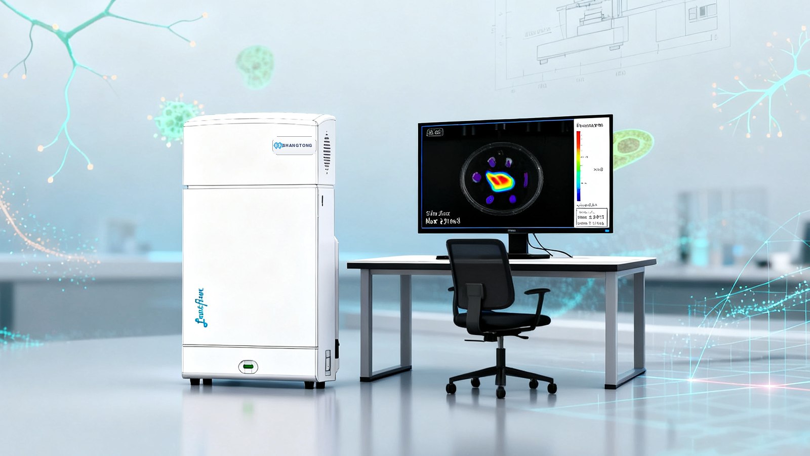
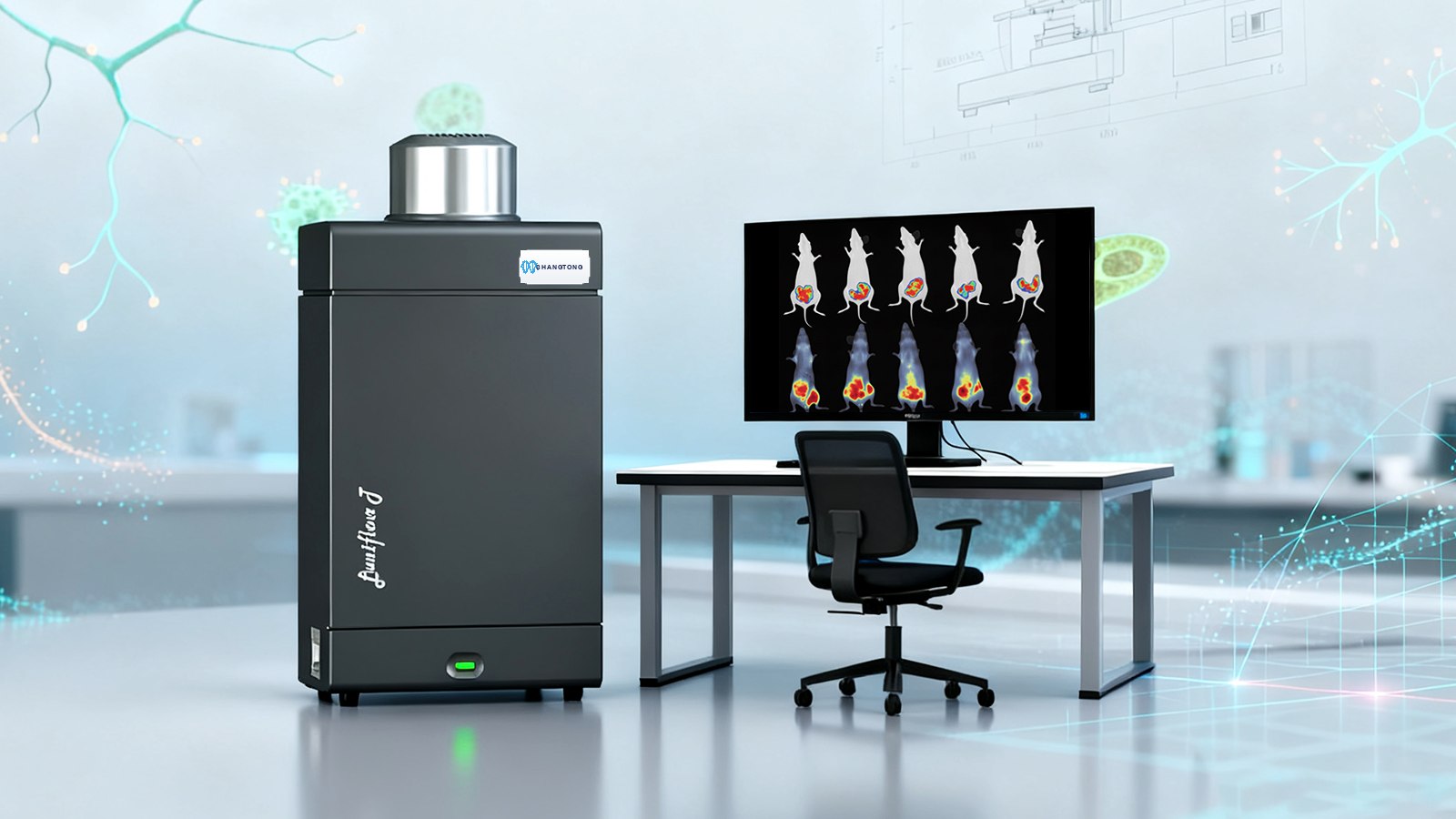
Product Gallery
Applications
Oncology Research
Non-invasive in vivo imaging of tumour growth, invasion and metastasis, with precise quantification of tumour volume and signal intensity.
Drug Development
Track the biodistribution, targeting, metabolism and clearance of fluorescent or radionuclide-labelled drugs in vivo.
Nanomaterial & Translational Medicine
Long-term in vivo tracing of nanomaterials and labelled cells to support translational research.
Technical Specifications
| Model | AVIS S I | AVIS TM 700 | AVIS TM 800 | AVIS T |
|---|---|---|---|---|
| Camera | 7.5 MP | 7.8 MP | 8.5 MP | 10.0 MP |
| Lens | F0.8 | F0.85 | F0.8 | F0.8 |
| Fluorescence System | 22-position LED, 22 included | 22-position LED, 22 included | 24-position LED, 24 included | 24-position LED, 24 included |
| Filter Wheel | 18-position, 8 included | 18-position, 10 included | 24-position, 10 included | 24-position, 10 included |
| Spectral Range | 400–900 nm | 400–900 nm | 400–900 nm | 400–900 nm |
| Max FOV | 20×20 cm | 25×25 cm | 25×25 cm | 26×26 cm |
How It Works
The platform the range is built on
LumiFluor AVIS is the reference configuration: deep-cooled detection, a calibrated optical path, motorised excitation and emission selection, and one software stack for acquisition and analysis. Every other model in the family inherits this chain and changes one variable — sensitivity, modality, spectral range or dimensionality.
Sensitivity is a chain, not a single number
Quantum efficiency, aperture, cooling and read noise each set a ceiling, and the weakest link decides the outcome. A back-illuminated sensor behind an F0.8 lens with −100 °C cooling and 16-bit digitisation is the combination that lets a small number of labelled cells be detected through tissue without binning the image into a blur.
Multimodality without re-alignment
Bioluminescence, fluorescence and brightfield frames share one optical path, so they overlay pixel-for-pixel. Optional X-ray, Cerenkov and upconversion modules extend the same chassis, which is why a laboratory can add a modality for the next project without re-validating its pipeline.
Choosing Within the AVIS Family
Start from the endpoint you must report, then pick the model that produces it with the least workflow overhead.
| Endpoint | Model | Why |
|---|---|---|
| Tumour burden, superficial, cohort scale | Faye A / T | Planar readout at the throughput a cohort needs |
| Very weak signals, multiplexed fluorescence | S 5 | Back-illuminated sensor plus F0.7 optics |
| Signal localisation in three dimensions | Spectrum / Spectrum 3D CT | Tomographic reconstruction |
| Bone reference and body composition | XRMS / X 6 | Dual-energy or switchable X-ray |
| NIR-II and full-spectrum panels | FS AVIS / NIR-II AVIS | InGaAs detection to 1700 nm |
Frequently Asked Questions
Which model should a new laboratory buy first?
Most groups start with a planar instrument because it covers reporter-gene efficacy work at cohort scale, and add a tomographic or X-ray model when a specific study demands it. If your first project already needs depth or bone reference, buy that capability up front rather than planning a second purchase.
How do I know whether my signal is strong enough?
Send us the reporter construct, the cell number and the implantation site. We reproduce a comparable model during the demonstration and show you the measured signal-to-noise at realistic exposures — a specification sheet cannot answer this question, an acquisition can.
What is included in a standard installation?
Optical alignment verification, dark-current and flat-field calibration, anaesthesia and stage setup, an operator walkthrough and a supervised first study, followed by remote sessions while your first results are reviewed.
How is the system serviced?
Preventive maintenance covers the detector cooling performance, optical transmission and stage calibration, with remote diagnostics in between. Consumables are limited to filters, lamps and anaesthesia parts, all of which are catalogued in the service plan.
Can data be analysed outside the supplied software?
Yes. Exports are 16-bit TIFF with metadata, and region measurements are exported as CSV, so ImageJ/Fiji, Python and R pipelines all work. We document the calibration factors so that values computed outside the software remain comparable with in-software results.
Related Reading
Request a Quote
Contact our team to configure the ideal in vivo imaging system for your research.
Contact Us